
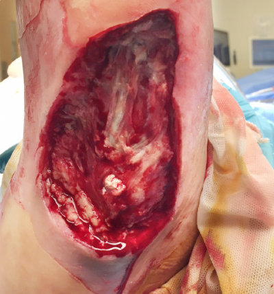

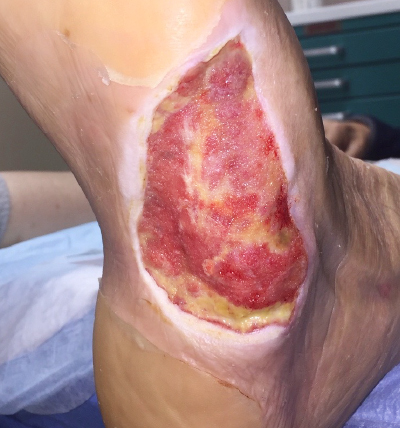
Initial Presentation
Week 0 – Post-debridement
- Intravenous Antibiotics
- Endoform Natural applied, plus a contact layer, under NPWT
- Wound Size: 9 cm x 6.5 cm x 2.5 cm
- Case Notes: Exposed tendon
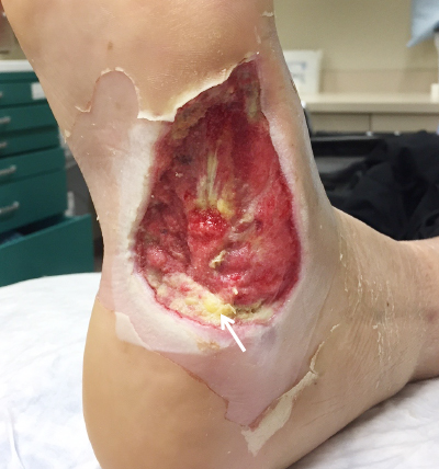
Week 1
- Endoform Natural and NPWT
- Beefy red granulation tissue
- Some residual Endoform in the wound bed (arrow)
- Evidence of epithelialization at the wound margins
- Wound Size: 8.2 cm x 5.8 cm x 0.4 cm
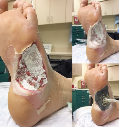
Week 1 - Application Tip
- Apply Endoform
- Cover with a non-contact layer
- Apply NPWT over the Endoform
Week 3
- Endoform Natural plus NPWT continued
- Endoform incorporating into wound bed
- Advancing epithelial tissue
- Wound Size: 8.5 cm x 5.2 cm x 0.3 cm
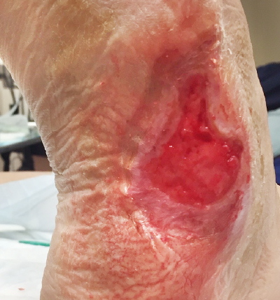
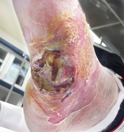
Week 17
- NPWT discontinued
- Endoform Natural reapplied, and foam dressing added under a TCC
- HBO
- Epithelial tissue covers half of the wound
- Wound Size: 4.3 cm x 3.2 cm x 0.1 cm
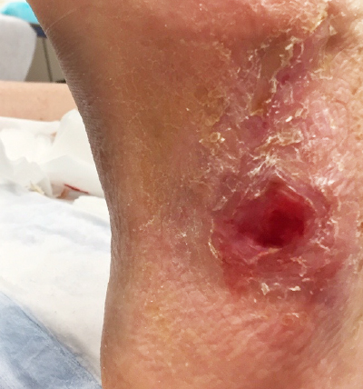
Week 24
- Endoform Natural reapplied, and foam dressing added under a TCC
- Wound size dramatically decreased
- Wound Size: 1 cm x 0.9 cm
Week 28
- 100% Re-epithelialization
- Wound Size: Healed